stethoscope supplier
Focus on Premium Stethoscope Customization
( Parts or Whole Set )

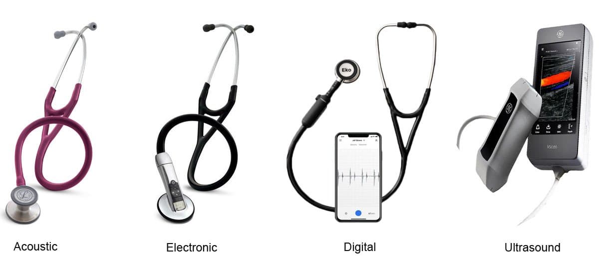
In today’s market, there are three general types of stethoscopes available to consumers: the traditional acoustic, amplifying, and digitizing. All of the electronic stethoscopes fall into two broad categories; amplifying and digitizing.

Stethoscope (from the Greek stethos-chest and skope-examination) is an acoustic medical device for auscultation, or listening to the internal sounds of a body. The stethoscope was invented in France in 1816 by Rene Laennec. It consisted of a wooden tube and was monoaural, which meant you could only hear through one ear when utilizing it. Prior to Laennec’s invention, physicians would perform “direct auscultation” to listen to patient’s internal sounds, which consisted of placing their ear directly on the patient’s body. In 1851 Arthur Leard invented the binaural stethoscope, which most closely resembled the traditional acoustic device as we know it today. The function of the acoustic stethoscope is attributed to three main components discussed further below. The components are:
Chest Piece
The acoustic stethoscope operates on the transmission of sound from the chest piece, via air-filled hollow tubes, to the listener’s ears. The chest piece usually consists of two parts that can be placed against the patient for sensing sound: the bell (hollow cup) and the diaphragm (disc). The bell transmits lower frequency sounds, while the diaphragm transmits higher frequency sounds. When the bell is placed on the patient, the vibrations of the skin directly produce acoustic pressure waves that travel to the listener’s ear. When the diaphragm is placed against the patient’s skin, body sounds vibrate the diaphragm, creating acoustic pressure waves which then travel to the listener’s ears. The human body then translates these pressure waves into sound and allows the listener to “hear” what they are examining.
With advances in acoustic stethoscope design, various companies have also come up with newer single-head chest piece designs, otherwise known as “dual frequency” or “tunable” diaphragms. These newer diaphragms have a one-sided chest piece and rely on user-applied pressure to change between bell and diaphragm modes. For low-frequency sounds (bell mode), light contact is used on the chest piece. The diaphragm membrane is then contained by a flexible surround that actually suspends it, allowing for the traditional functionality and sound transfer of the bell. For high-frequency sounds (diaphragm mode), firm contact pressure is used on the chest piece. By pressing on the chest piece, the diaphragm membrane moves inward until it reaches an internal ring. The ring simply restricts the diaphragm membrane’s movement and allows for the traditional functionality and sound transfer mechanism of the diaphragm.
Stethoscope tubing is the air-filled hollow tubing that allows sound energy to be transmitted from the chest piece to the clinician. Tubing can vary by manufacturer and comes in different varieties; many are now latex free. There are single-lumen tubes which are usually split into two tubes at the ear pieces, bi-lumen tubes that consist of two internal lumen incorporated into a single lumen design, and tubing systems that consist of two separate tubes that are directly connected from chest piece to each ear piece. Each type of stethoscope tubing has its own resonant frequency which depends on the length and internal width of the tubing, similar to a pipe organ. (Finkelstein, 2008) Thicker tubing can better insulate the sound being transmitted against outside distortion. An increase in the length of the tubing will decrease the pressure at the end of the tubing as a result of frictional and other internal forces. The overall change in length between most stethoscope tubing is relatively small. Therefore, the decrease in acoustic pressure is generally not thought to be detectable by the human ear. As tubing length increases, resonant frequency decreases and sounds have greater potential to be attenuated. Tubing length tends to come down to preference, but is usually between 18-26 inches.
Ear Pieces
When a Clinician places the stethoscope chest piece on a patient, they are closing a circuit that allows sound energy to travel internally from the patient, through a conductor (the stethoscope), and to the listener. In this type of circuit, a break or air leak in the circuitry may result in decreased sound quality or completely block the transmission of sound. The physical connection that is made between the ear tips and the human ear is very important. The insertion pressure, ear tip seal, and ear tip insertion angle are all variables that can affect this connection. Some manufactures offer hard and soft sealing ear tips, various sizes of ear tips to accommodate ear canal size. These give the user the ability to adjust the insertion pressure and angle by manually manipulating the head set. When using an acoustic stethoscope, it is important that the ear pieces angle forward to fit most naturally into the external ear opening.
One problem with acoustic stethoscopes is that the sound level is extremely low. Electronic stethoscopes utilize advanced technology to overcome these low sound levels by electronically amplifying body sounds. Electronic stethoscopes require conversion of acoustic sound waves obtained through the chest piece into electronic signals which are then transmitted through uniquely designed circuitry and processed for optimal listening. The circuitry consists of components that allow the energy to be amplified and optimized for listening at various frequencies. The circuitry also allows the sound energy to be digitized, encoded and decoded, to have the ambient noise reduced or eliminated, and sent through speakers or headphones.
Unlike acoustic stethoscopes, which are all based on the same physics, transducers in electronic stethoscopes vary widely. The simplest and least effective method of sound detection is achieved by placing a microphone in the chest piece. This method suffers from ambient noise interference. Another method comprises placement of a piezoelectric crystal at the head of a metal shaft, the bottom of the shaft making contact with a diaphragm. Some manufacturers use a piezoelectric crystal placed within foam behind a thick rubber-like diaphragm. Another manufacturer uses an electromagnetic diaphragm with a conductive inner surface to form a capacitive sensor. This diaphragm responds to sound waves identically to a conventional acoustic stethoscope, with changes in an electric field replacing changes in air pressure. This preserves the sound of an acoustic stethoscope with the benefits of amplification. No matter what type of sophisticated circuitry or transducer is utilized, practitioners need to be prepared for a difference in the sound quality between acoustic and electronic stethoscopes. Clinicians will automatically recognize an “electronic” quality to even the best sounds obtained from the electronic stethoscopes currently on the market. Utilizing electronic stethoscopes for auscultation definitely takes getting used to, but with repeated exposure any practitioner can learn to appreciate the sound obtained with an electronic stethoscope.
The fact that sounds are transmitted electronically allows electronic stethoscopes to offer features such as audio or serial data output, wireless transmission, and recording of sound clips.
Amplifying Stethoscopes
The majority of the electronic stethoscopes on the market have an audio output signal that, through the use of a stereo and/or mono cable connection, can allow the audio output collected by the stethoscope to be transmitted real time to an accompanying software application. The accompanying software may employ various algorithms that are developed for interpretation and diagnosis of the audio output obtained from the electronic stethoscopes. Audio output processed through various accompanying software packages can be saved as files and then transmitted via email and various other communication methods for asynchronous assessment and diagnosis. Some electronic stethoscopes that have audio data output options can also be hooked up to a videoconferencing unit’s audio input, which would enable them to send the sound data real time over a network connection to another videoconferencing endpoint. The videoconferencing units would act as a decoders and encoders of the sound data, allowing it to be transmitted over a network. In this type of setup, the user needs to be mindful of the fact that generally the sound data can only be sent between like brands of videoconferencing units and sometimes as specific as like models of like brands. Depending on the speakers at the other end of the videoconference, some of the frequencies obtained may not be discernable, and the audio output quality has the potential to suffer. This adjunct to the videoconferencing session may also utilize additional bandwidth, causing the quality of the video session to suffer as well. Additionally, if the video participants wish to speak to each other during the session, the users may have to set up a means to also allow this communication to occur because only one type of audio transmission may be allowed at a time. Some electronic stethoscopes allow transmission of data through a wireless or Bluetooth interface to other devices. Most of the wireless electronic stethoscopes require dongles (Bluetooth receivers) to transmit data wirelessly. Wireless and Bluetooth data communication strategies utilize more recent technologies. However, this form of data transmission may be a limiting factor in compatibility with devices other than computers; videoconferencing units for example.
Only one stethoscope currently on the market allows for onboard recording and playback directly into the physical portion of the stethoscope. Any electronic stethoscope that has data transmission and a software interface will allow for recording of clips that are not stored locally on the device. Some of the electronic stethoscopes offer visual output of the heart rate and EKG waves detected directly on the device and all allow for visual output of the sound waves collected in conjunction with their accompanying software packages.
Digitizing Stethoscopes
A few of the electronic stethoscopes on the market are called “digitizing stethoscopes” because they convert the audio sound to a digital signal. These stethoscopes can transmit serialized audio data that can be shared real time (synchronously) and/or in a store and forward fashion (asynchronously). These units work by detecting sound through the electronic stethoscope sensor, converting that sound energy to electricity and running it through circuitry which can amplify it, filter it by frequency, and finally convert the data from analog to a digital. Click here to learn more about digital to analog conversion. Basically, the units themselves are codecs that digitally encode and decode data so it can be transmitted over a network. After the data is digitized, it can be sent through a videoconferencing unit through its RS32 input across a network or through computer software with a serial TCP/IP converter and across a network. One stethoscope can transmit the digital data using USB to store-and-forward software. Both types of data transmission require that there is a corresponding electronic stethoscope unit on the other side that will convert the digital signal back to an analog signal, amplify the sound, filter the sound frequency, and transmit the sound energy to the human ear.